LLU Fertility IVF Lab
Accredited andrology & IVF labs
Learn about Loma Linda University Center for Fertility’s accredited andrology & IVF labs, including our licensed tissue bank, licensed lab testing, and accredited ambulatory surgery center.
Embryology, IVF & genetic testing services
The embryology lab at the Loma Linda University Center for Fertility & IVF is responsible for identifying and processing patient oocytes for fertilization, embryo culture and selection, and for cryopreservation and storage of embryos. In addition, the IVF lab also processes embryo biopsies for preimplantation genetic diagnosis (PGT-M).
Our IVF lab services include:
- Female egg (oocyte) retrieval, incubation, evaluation and fertilization
- Unique incubator chambers for each patient’s embryos
- PGT-M and screening
- Selecting optimal embryos for day 3 or 5 blastocyst transfer
- Embryo transfer lab assistance, including:
- Acupuncture
- The application of embryo glue to assist in embryo implantation during the transfer
- Vitrification for freezing eggs or embryos for use in a later IVF treatment (cycle), post-treatment storage or for fertility preservation.
Egg retrieval
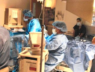 Under ultrasound guidance, the patient’s oocytes (eggs) are removed from the ovaries and placed into pre-warmed culture tubes. The oocytes are bathed in a mixture of follicular fluid and culture medium.
Egg evaluation & fertilization
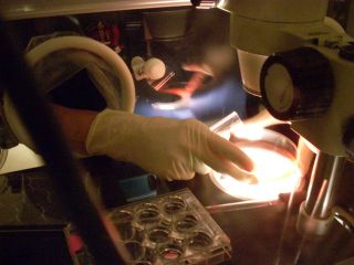
The oocytes are identified using a dissecting microscope and categorized as mature, immature or atretic (dying). Each oocyte is washed clean of cellular debris and incubated in a separate tissue culture medium rich in nutrients.
The mature oocyte is then ready to be fertilized with washed sperm cells (conventional IVF). Sperm are added into a special culture with the egg for natural fertilization to occur.
Egg fertilization with ICSI
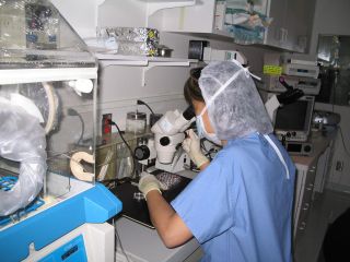
A mature oocyte can also be fertilized by injecting a single sperm into the egg (ICSI). Before this can take place, the layers of cumulus cells surrounding each oocyte have to be removed to facilitate the procedure. The cumulus removal process is performed using another microscope with a warm stage.
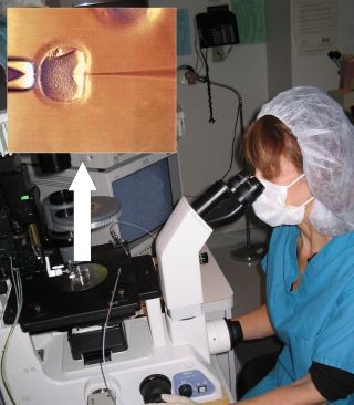
The oocyte is held in place by a large pipet that has a gentle suction pressure. A fine needle pipet containing a single washed sperm is injected inside the oocyte. The oocyte is released and placed back into the tissue culture well with culture medium. Then the lab incubates all the injected oocytes overnight in incubators.
Confirming fertilization
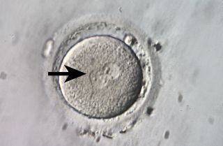
The day following oocyte retrieval and fertilization, each oocyte is examined under the inverted microscope to see if fertilization was successful. This is usually confirmed by the presence of two pronuclei near the center of the oocyte (where the arrow is pointing in this photo).
Unfertilized oocytes do not have any pronuclei present. The physician and patient are informed of the number of fertilized oocytes, which are then incubated for another day and re-examined for development.
Embryo development
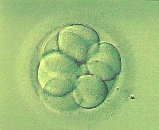
The faster developing embryos reach the 8-cell stage as shown in this photo by day 3 of development. This is usually a good sign that the embryo will continue to develop and be a good option for transfer. Not all the individual cells will be round and even, and they may be larger or smaller in size. However, as long as a few of the cells are normal, the embryo has good developmental potential.
Embryo genetic testing
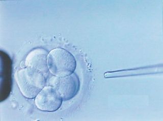
When the patient desires PGT-M ensure the genetic health of each embryo, two of the embryo’s eight cells are removed using a pipet. The extracted cells are processed and analyzed. The patient obtains the PGT-M results in less than 24 hours after the embryo biopsy.
Blastocyst stage: selecting an embryo for transfer
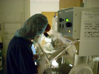
Five days after oocyte retrieval, the embryo has grown to the blastocyst stage. The blastocysts that have the best shape and development are selected for embryo transfer.
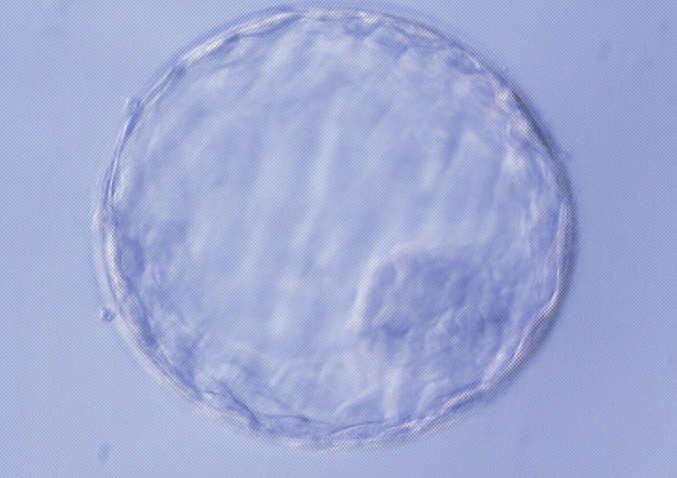
Some selection criteria include the appearance of blastocyst expansion, the presence of an inner fluid-filled space called the blastocele, the tightness of the cell layers, and the thickness of the zona pellucida (the egg’s outer shell). The remaining unused embryos are frozen and stored in accordance with the wishes of the patient.
Embryo transfer
The embryo selected for transfer is loaded into a plastic embryo transfer tube (catheter). The physician then guides the end of the catheter into the patient’s uterus through the cervical canal under ultrasound guidance. The embryo is gently placed in the uterus and the catheter is removed.
The patient is encouraged to rest for a few minutes and is given further instructions after the embryo transfer. A blood test is performed two weeks after the transfer to confirm pregnancy.
